")
26 min read
5137 words
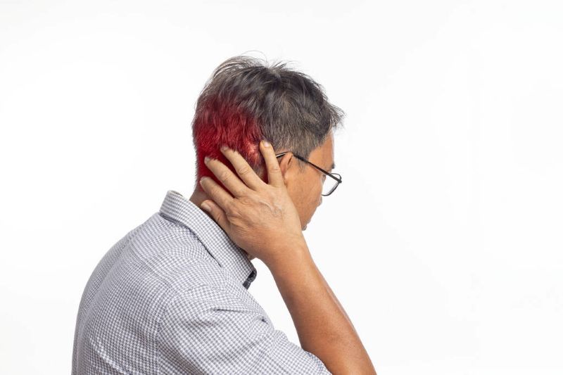
Imagine a sharp, radiating pain shooting down your arm, or a persistent tingle in your fingers that just won’t go away. Perhaps you’ve noticed a subtle weakness when trying to lift something, or a dull ache in your neck that seems to have a mind of its own. These aren’t just annoying discomforts; they could be the tell-tale signs of a “nerve pinch” – a common yet often debilitating condition known medically as cervical radiculopathy. And when we talk about the neck, the C5, C6, and C7 levels are often the primary culprits in this intricate dance of pain and dysfunction. Your neck, or cervical spine, is a marvel of engineering, allowing an incredible range of motion while simultaneously housing and protecting your spinal cord and the vital nerve roots that branch out to control your arms, hands, and upper body. When one of these delicate nerve roots gets compressed or irritated, often by a herniated disc, bone spur, or narrowed spinal canal, the consequences can be far-reaching, transforming daily tasks into painful struggles. This blog post will serve as your comprehensive guide to understanding a pinched nerve affecting the C5, C6, and C7 segments of your neck. We’ll delve into the anatomy that makes these areas vulnerable, explore the myriad causes behind nerve compression, dissect the distinct symptoms you might experience, and chart a path through the diagnostic process. Most importantly, we’ll illuminate the full spectrum of treatment options, from conservative therapies that often bring significant relief to the more advanced surgical interventions that can restore function and quality of life. Our aim is to empower you with knowledge, helping you navigate the complexities of this condition and take proactive steps towards healing and well-being.
Understanding the Cervical Spine: The Foundation of Your Neck’s Mobility and Sensation
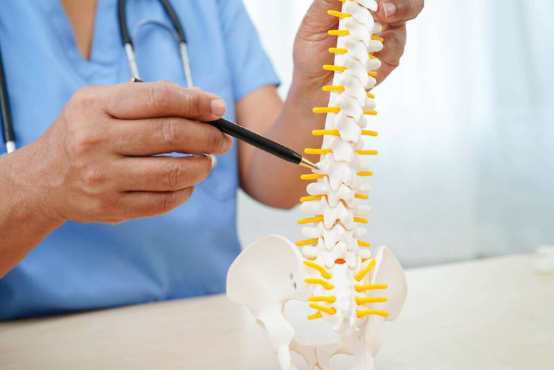
Before we dive into the specifics of a nerve pinch, it’s crucial to appreciate the sophisticated structure of your cervical spine. Comprising seven individual vertebrae, labeled C1 through C7, this section of your spine starts at the base of your skull and extends down to your upper back. Each vertebra is separated by an intervertebral disc – a soft, gel-filled cushion that acts as a shock absorber and allows for flexible movement.
Beyond providing structural support, the cervical spine forms a protective tunnel for the spinal cord, the main highway of your nervous system. At each level of the cervical spine, a pair of spinal nerve roots branches off the spinal cord, exiting through small openings called foramina (plural of foramen) on either side of the vertebrae. These nerve roots are like the electrical wires that carry signals: motor commands from the brain to your muscles, allowing movement, and sensory information from your skin, muscles, and joints back to the brain, enabling you to feel touch, temperature, and pain.
The C5, C6, and C7 nerve roots are particularly critical because they supply sensation and motor function to large areas of your shoulder, arm, and hand. For instance:
- The C5 nerve root primarily controls the deltoid muscle (shoulder abduction) and biceps (elbow flexion), while providing sensation to the outer part of your shoulder and upper arm.
- The C6 nerve root plays a significant role in wrist extension and elbow flexion through the biceps and brachioradialis muscles, and gives sensation to the thumb and index finger.
- The C7 nerve root is essential for elbow extension (triceps), wrist flexion, and finger extension, with sensory innervation to the middle finger.
The cervical spine is the most mobile part of your spinal column, which is both a blessing and a curse. This flexibility allows you to turn, tilt, and nod your head, but it also makes the discs and joints susceptible to wear and tear, injury, and degenerative changes over time. The constant motion and the transition zones within the spine, particularly around the lower cervical segments (C5-C7), make these areas highly vulnerable to the very conditions that lead to nerve compression. Understanding this fundamental anatomy is the first step in comprehending why a “pinch” at these specific levels can lead to such distinct and often debilitating symptoms throughout your upper extremity.
What Exactly is a “Nerve Pinch” (Cervical Radiculopathy)?
The term “nerve pinch” is a colloquial but accurate description of a medical condition known as cervical radiculopathy. At its core, cervical radiculopathy refers to the compression or irritation of a nerve root as it exits the spinal cord in the cervical spine (neck region). Think of it like a garden hose: when it’s kinked or something is pressing on it, the water flow is restricted. Similarly, when a nerve root is “pinched,” the normal flow of nerve signals is disrupted, leading to a cascade of symptoms that can range from mild discomfort to severe pain, numbness, tingling, and even muscle weakness in the areas supplied by that specific nerve.
It’s crucial to differentiate cervical radiculopathy from general neck pain or a simple muscle strain. While a muscle strain might cause localized stiffness and soreness in the neck, a nerve pinch typically produces symptoms that radiate away from the neck, down into the shoulder, arm, hand, or fingers. This radiating pattern is a hallmark of nerve involvement and helps clinicians pinpoint the affected nerve root.
Unlike temporary muscle fatigue, a nerve pinch represents a structural issue where something is physically encroaching upon the nerve. This impingement can cause inflammation, swelling, and disruption of the nerve’s ability to transmit signals effectively. The severity of symptoms often correlates with the degree of compression and inflammation.
The precise location of the pinch (which specific nerve root out of C5, C6, or C7) dictates the exact pattern of symptoms. Since each nerve root has a designated pathway – a dermatome for sensation and a myotome for muscle function – the doctor can often tell which nerve is affected based on where you feel pain, numbness, or weakness. This anatomical specificity is why understanding the C5-C7 segments is so vital, as these are the most commonly affected nerve roots leading to arm and hand symptoms. When a nerve is truly “pinched,” it’s a signal from your body that something is out of alignment or encroaching upon a critical communication pathway, demanding attention and appropriate intervention.
Causes of C5-C7 Nerve Pinches: Why Your Nerves Are Under Pressure

Nerve compression at the C5, C6, or C7 levels stems from a variety of structural changes and degenerative processes within the cervical spine. While some causes are acute and sudden, many are the result of cumulative wear and tear over time. Understanding these underlying mechanisms is key to effective diagnosis and treatment.
1. Herniated Disc (Disc Bulge or Rupture)
This is one of the most common acute causes of a nerve pinch. Each intervertebral disc consists of a tough outer fibrous ring (annulus fibrosus) and a soft, gel-like inner core (nucleus pulposus). A herniated disc occurs when the outer ring tears, allowing the inner gel-like material to push outward or even rupture.
- Mechanism: When the disc material pushes out, it can directly press against a nearby nerve root as it exits the spinal canal through the neural foramen. The extruded material also releases inflammatory chemicals, further irritating the nerve.
- How it happens: Often caused by a sudden, forceful movement, lifting heavy objects incorrectly, whiplash injury, or repetitive strain. It can also occur gradually due to disc degeneration, where the disc weakens over time, making it more susceptible to herniation.
2. Spinal Stenosis
Spinal stenosis refers to the narrowing of the spinal canal (the central canal where the spinal cord runs) or the neural foramen (the smaller openings through which nerve roots exit).
- Mechanism: This narrowing reduces the space available for the spinal cord and nerve roots, leading to compression.
- Causes:
- Bone Spurs (Osteophytes): As discs degenerate and vertebrae rub against each other, the body attempts to stabilize the spine by growing extra bone. These bone spurs can directly impinge on nerve roots or the spinal cord.
- Thickened Ligaments: The ligaments that support the spine can thicken and stiffen with age, encroaching on the neural structures.
- Disc Bulging/Herniation: While distinct, chronic disc issues often contribute to stenosis.
3. Degenerative Disc Disease (DDD)
DDD is not strictly a disease but rather a natural, age-related process of wear and tear on the intervertebral discs.
- Mechanism: Over time, discs lose their water content, becoming less pliable and thinner. This loss of disc height can bring the vertebrae closer together, narrowing the neural foramina and making nerve compression more likely. Degenerated discs are also more prone to bulging and herniation, and often trigger the formation of bone spurs.
- Contribution to Nerve Pinch: DDD often acts as a precursor or contributing factor to both herniated discs and spinal stenosis, creating an environment where nerve roots are vulnerable.
Less Common Causes:
- Trauma: Severe injuries like fractures or dislocations can directly damage and compress nerve roots.
- Tumors: Although rare, growths within or near the spinal canal can compress nerve roots.
- Infections: Spinal infections can cause inflammation and structural changes leading to compression.
- Cysts: Fluid-filled sacs can sometimes develop and press on nerves.
Contributing Factors:
Beyond these direct causes, several factors can increase one’s risk of developing a nerve pinch:
- Poor Posture: Chronic slumping, forward head posture (“tech neck”), or improper sleeping positions can put undue stress on the cervical spine and accelerate disc degeneration.
- Repetitive Strain: Certain occupations or activities involving prolonged neck flexion, rotation, or heavy lifting can contribute to disc and joint wear.
- Genetics: Some individuals may be genetically predisposed to conditions like DDD.
- Smoking: Nicotine impairs blood flow to the discs, accelerating their degeneration.
- Obesity: While less directly impactful on the cervical spine than the lumbar spine, excess weight can contribute to overall spinal stress.
Understanding the interplay of these factors is crucial for prevention and for developing a comprehensive treatment strategy that addresses not just the symptoms but also the underlying causes of a C5-C7 nerve pinch.
The Distinctive Symptoms of C5 to C7 Nerve Compression
A nerve pinch in the cervical spine doesn’t just cause generic neck pain; it produces a constellation of symptoms that are often specific to the particular nerve root being compressed. This specificity is incredibly useful for diagnosis, serving as a roadmap for healthcare professionals to pinpoint the exact level of impingement. While there can be some overlap, recognizing the unique patterns associated with C5, C6, and C7 compression is key to understanding your own experience.
General Symptoms of Cervical Radiculopathy:
Before dissecting the individual nerve roots, let’s look at the broad categories of symptoms:
- Neck Pain: Often localized, but can radiate to the shoulder blade or upper back. It might be dull, aching, sharp, or throbbing.
- Radicular Pain: This is the hallmark symptom – pain that radiates from the neck, down the arm, forearm, and into the hand or fingers. It often follows a narrow band, distinguishing it from more diffuse muscle pain. This pain can be burning, electric-shock-like, or deep and aching.
- Paresthesia (Numbness or Tingling): A sensation of “pins and needles,” decreased feeling, or complete numbness in the affected dermatome (area of skin supplied by a single nerve root).
- Weakness (Motor Deficit): Difficulty with specific movements or loss of strength in muscles supplied by the compressed nerve root (myotome). This can manifest as difficulty grasping objects, lifting the arm, or extending the elbow.
- Diminished Reflexes: Changes or absence of deep tendon reflexes (e.g., biceps, brachioradialis, triceps reflexes).
- Increased Pain with Neck Movement: Certain neck positions or movements, particularly extending the neck or turning the head towards the affected side (Spurling’s maneuver), can exacerbate symptoms.
Now, let’s break down the specific symptom patterns for C5, C6, and C7 nerve root compression:
C5 Nerve Root Compression Symptoms:
When the C5 nerve root is pinched, the symptoms primarily affect the upper shoulder and arm.
- Pain Distribution: Often felt in the side of the neck, radiating across the top of the shoulder to the outer part of the upper arm (deltoid region).
- Numbness/Tingling (Dermatome): Typically experienced on the outer aspect of the shoulder and upper arm.
- Weakness (Myotome): Weakness in the deltoid muscle (making it difficult to lift the arm away from the body, known as shoulder abduction) and sometimes the biceps muscle (difficulty bending the elbow). A feeling of “giving way” in the shoulder can occur.
- Reflex Loss: The biceps reflex (a tap on the biceps tendon near the elbow) may be diminished or absent.
C6 Nerve Root Compression Symptoms:
Compression of the C6 nerve root is one of the most common types of cervical radiculopathy, affecting the lateral forearm, thumb, and index finger.
- Pain Distribution: Pain commonly radiates down the side of the arm, along the forearm, and into the thumb and index finger. It can also be felt in the neck and shoulder blade area.
- Numbness/Tingling (Dermatome): Predominantly felt in the thumb and index finger, and along the radial (thumb side) aspect of the forearm.
- Weakness (Myotome): Weakness in the biceps muscle (difficulty with elbow flexion) and wrist extensors (difficulty bending the wrist upwards). You might notice difficulty with tasks requiring grip or lifting.
- Reflex Loss: The brachioradialis reflex (a tap on the forearm bone near the wrist) may be diminished or absent. The biceps reflex can also be affected.
C7 Nerve Root Compression Symptoms:
The C7 nerve root is also very frequently affected, leading to symptoms that extend to the middle finger and the back of the arm.
- Pain Distribution: Pain often travels down the back of the arm (triceps area), along the forearm, and into the middle finger (and sometimes the index and ring fingers).
- Numbness/Tingling (Dermatome): Primarily experienced in the middle finger, and sometimes adjacent fingers.
- Weakness (Myotome): Significant weakness in the triceps muscle (difficulty straightening the elbow, pushing, or “pushing away” movements). Weakness can also affect wrist flexors and finger extensors, making it hard to grip or extend the fingers.
- Reflex Loss: The triceps reflex (a tap on the triceps tendon just above the elbow) may be diminished or absent.
When to Seek Immediate Medical Attention:
While most nerve pinches are not medical emergencies, certain symptoms warrant immediate evaluation by a healthcare professional:
- Progressive Weakness: If weakness rapidly worsens or affects multiple muscle groups.
- Bowel or Bladder Dysfunction: New onset of difficulty controlling urination or bowel movements, which could indicate spinal cord compression (myelopathy).
- Difficulty Walking or Balance Issues: Suggests potential spinal cord involvement.
- Bilateral Symptoms: Symptoms affecting both arms or hands.
- Severe, Unremitting Pain: Pain that is not relieved by rest or over-the-counter medication.
Understanding these specific symptom patterns is the first step towards an accurate diagnosis and, consequently, an effective treatment plan for your C5-C7 nerve pinch.
Diagnosis: Unraveling the Mystery of Your Neck Pain
Accurately diagnosing a C5-C7 nerve pinch requires a comprehensive approach, combining a thorough medical history, a detailed physical and neurological examination, and often, advanced imaging studies. The goal is not only to confirm the presence of nerve compression but also to identify its exact location and underlying cause.
1. Clinical Examination: The Foundation
The diagnostic process typically begins with a visit to a healthcare provider, such as a general practitioner, chiropractor, physical therapist, or spine specialist (neurologist, orthopedist, neurosurgeon).
- History Taking: This is a crucial first step. Your doctor will ask detailed questions about your symptoms:
- When did the pain start? Was there an injury?
- What exactly does the pain feel like (sharp, dull, burning, tingling, electric)?
- Where does the pain radiate? (This helps pinpoint the nerve root).
- What makes the pain better or worse? (e.g., neck movements, arm positions, rest).
- Have you experienced any numbness, tingling, or weakness? If so, where?
- Do you have any other symptoms like balance issues or bowel/bladder changes?
- What treatments have you tried so far?
- Your occupation, hobbies, and overall health history are also important.
- Physical Examination: This involves a series of tests to assess your neck and neurological function:
- Observation: Assessing posture, alignment of the head and neck.
- Palpation: Gently feeling the neck muscles and spine for tenderness, spasms, or abnormalities.
- Range of Motion: Evaluating how far you can move your neck (flexion, extension, rotation, lateral bending). Limited or painful movement can indicate an issue.
- Neurological Examination: This is critical for identifying nerve root involvement:
- Sensory Testing: Light touch, pinprick, and temperature sensation are tested in various areas of the arm and hand to map out dermatomal deficits (e.g., C6 dermatome for thumb/index finger).
- Motor Strength Testing: You’ll be asked to perform specific movements against resistance (e.g., shoulder shrug, bicep curl, tricep extension, wrist extension/flexion, grip strength). This helps identify myotomal weakness (e.g., C7 myotome for triceps).
- Reflex Testing: Deep tendon reflexes (biceps, brachioradialis, triceps) are tested. Diminished or absent reflexes are strong indicators of nerve root compression at specific levels.
- Special Provocative Tests:
- Spurling’s Maneuver: The doctor extends and rotates your neck to the affected side and applies gentle downward pressure. If this reproduces your radiating arm pain, it strongly suggests nerve root compression.
- Distraction Test: Gently lifting the head (distracting the cervical spine) can sometimes relieve nerve compression symptoms, further supporting the diagnosis.
2. Imaging Studies: Visualizing the Problem
While the clinical exam provides strong clues, imaging helps confirm the diagnosis, show the anatomical cause, and rule out other conditions.
- X-rays:
- What it shows: Primarily bone structures. Can reveal spinal alignment, degenerative changes (bone spurs, disc space narrowing), and fractures.
- Limitations: Does not show soft tissues like discs, nerve roots, or the spinal cord. Useful as an initial screen.
- MRI (Magnetic Resonance Imaging):
- Gold standard for soft tissues: This is often the most informative imaging study for cervical radiculopathy.
- What it shows: Provides detailed images of the spinal cord, nerve roots, intervertebral discs, and ligaments. It can clearly visualize herniated discs, spinal stenosis (from disc bulging, bone spurs, or thickened ligaments), and inflammation.
- Why it’s important: Crucial for identifying the precise level and nature of nerve root compression.
- CT Scan (Computed Tomography):
- What it shows: Excellent for detailed bone imaging, often better than X-rays. Can clearly show bone spurs, osteophytes, and the bony dimensions of the spinal canal and foramina.
- When it’s used: Often used if an MRI is contraindicated (e.g., pacemakers, certain metal implants) or when more detailed bone information is needed, especially in pre-surgical planning. Sometimes combined with myelography (CT myelogram) where dye is injected into the spinal canal to highlight nerve compression.
- EMG/NCS (Electromyography/Nerve Conduction Studies):
- What it shows: These electrodiagnostic tests assess the electrical activity of muscles and the speed of nerve signal conduction.
- When it’s used:
- To confirm nerve root compression and assess its severity.
- To differentiate cervical radiculopathy from other conditions that can cause similar arm symptoms, such as carpal tunnel syndrome, ulnar neuropathy, or brachial plexopathy (peripheral nerve entrapments).
- To determine if there’s ongoing nerve damage.
By combining the detailed information from your symptoms and physical examination with the visual evidence from imaging and the functional assessment from electrodiagnostic tests, your healthcare team can develop an accurate diagnosis and then formulate the most appropriate and effective treatment plan for your specific C5-C7 nerve pinch.
A Comprehensive Approach to Healing – Treatment Options for C5-C7 Nerve Pinches

Dealing with a C5-C7 nerve pinch can be challenging, but the good news is that a wide array of treatment options are available, with the vast majority of patients finding relief through conservative, non-surgical methods. The approach is typically stepped, starting with the least invasive options and progressing to more aggressive interventions only if necessary. The primary goals of treatment are to reduce pain, decrease inflammation, decompress the affected nerve root, and restore function.
1. Conservative Management (First Line of Treatment)
For most individuals with cervical radiculopathy, conservative treatments are the cornerstone of recovery and are often successful within 6-12 weeks.
- Rest and Activity Modification:
- Principle: Avoid activities and positions that aggravate your symptoms. This doesn’t mean complete bed rest, but rather modifying daily tasks to reduce stress on your neck.
- Application: Temporarily avoiding heavy lifting, overhead activities, prolonged sitting with poor posture, or specific neck movements that trigger pain. Learning proper body mechanics is key.
- Medications:
- NSAIDs (Non-Steroidal Anti-inflammatory Drugs): Over-the-counter options like ibuprofen (Advil, Motrin) or naproxen (Aleve) can reduce pain and inflammation. Prescription-strength NSAIDs may also be used.
- Muscle Relaxants: Medications like cyclobenzaprine (Flexeril) or tizanidine (Zanaflex) can help alleviate muscle spasms that often accompany nerve irritation. Used short-term due to potential side effects like drowsiness.
- Oral Corticosteroids: A short course of corticosteroids (e.g., prednisone) can be prescribed to rapidly reduce severe inflammation around the nerve root, offering quick relief. However, they are not a long-term solution.
- Neuropathic Pain Medications: For persistent nerve pain, medications specifically designed to calm overactive nerve signals, such as gabapentin (Neurontin) or pregabalin (Lyrica), may be prescribed. These often take time to become effective.
- Physical Therapy (PT):
- Goals: Reduce pain, improve neck range of motion, strengthen supporting muscles, improve posture, and educate on self-management.
- Techniques:
- Manual Therapy: Hands-on techniques by a therapist, including gentle mobilization of the neck joints, soft tissue massage to reduce muscle tension, and targeted stretching.
- Therapeutic Exercises: A tailored program of exercises to:
- Stretch: Improve flexibility of tight neck and shoulder muscles.
- Strengthen: Build endurance and strength in the deep neck flexors, scapular stabilizers, and core muscles to support the cervical spine.
- Postural Correction: Exercises and awareness training to correct forward head posture and promote proper spinal alignment.
- Traction: Gentle, controlled pulling of the head (manual or mechanical) can help create space between vertebrae, temporarily decompressing the nerve root.
- Modalities: Heat, ice, ultrasound, or TENS (Transcutaneous Electrical Nerve Stimulation) may be used to manage pain and inflammation.
- Ergonomic Advice: Guidance on setting up your workstation, sleeping positions, and daily activities to minimize stress on your neck.
- Cervical Collar:
- Use: A soft cervical collar may be recommended for short-term use to immobilize the neck, reduce muscle spasms, and provide temporary pain relief, especially during flare-ups.
- Caution: Prolonged use is generally discouraged as it can lead to muscle weakening and stiffness.
- Injections:
- Epidural Steroid Injections (ESIs): A powerful anti-inflammatory corticosteroid, often combined with a local anesthetic, is injected into the epidural space surrounding the inflamed nerve root. This can provide significant, though often temporary, pain relief by reducing inflammation directly at the source. They are typically performed under X-ray guidance (fluoroscopy) for precision.
- Nerve Block Injections: Similar to ESIs, these target specific nerve roots with local anesthetic and steroids to block pain signals and reduce inflammation.
- Trigger Point Injections: If muscle spasms are a significant component of your pain, injections into specific trigger points can help release tension.
2. Surgical Intervention (When Conservative Fails or Neurological Deficits Worsen)
Surgery is typically considered a last resort for cervical radiculopathy, reserved for cases where extensive conservative treatment (usually 6-12 weeks or longer) has failed to provide sufficient relief, or if there are signs of progressive neurological deficits (such as increasing weakness, numbness, or loss of function) or spinal cord compression (myelopathy).
- Indications for Surgery:
- Intractable pain that severely impacts quality of life despite conservative efforts.
- Progressive neurological deficit (e.g., worsening arm weakness).
- Evidence of spinal cord compression (myelopathy), often indicated by balance issues, gait disturbances, or bowel/bladder changes.
- Clear anatomical compression identified on imaging that correlates with symptoms.
- Common Surgical Procedures:
- Anterior Cervical Discectomy and Fusion (ACDF): This is one of the most common and historically successful surgeries for cervical radiculopathy caused by disc herniation or stenosis.
- Procedure: An incision is made at the front of the neck. The surgeon removes the damaged intervertebral disc and any bone spurs that are compressing the nerve root and/or spinal cord. To maintain spinal stability, the space left by the disc is filled with a bone graft or a cage, and the vertebrae above and below are permanently fused together using a plate and screws.
- Benefits: Highly effective at relieving nerve compression and stabilizing the spine.
- Considerations: Loss of motion at the fused segment; potential for “adjacent segment disease” (increased stress on the discs above and below the fusion over time).
- Posterior Cervical Laminoforaminotomy:
- Procedure: An incision is made at the back of the neck. A small window is created in the lamina (the bony arch of the vertebra) and/or the foramen (nerve root exit hole) to relieve pressure on the nerve root, often by removing bone spurs or parts of the disc. This procedure aims to decompress the nerve without fusing the vertebrae, thus preserving motion.
- Benefits: Preserves motion, often faster recovery than fusion in some cases, less risk of adjacent segment disease.
- Considerations: Only suitable for nerve root compression (not spinal cord compression) and when the problem is lateral (to the side) rather than central.
- Artificial Disc Replacement (ADR):
- Procedure: Similar to ACDF, the damaged disc is removed, but instead of fusion, an artificial disc device is inserted into the disc space.
- Benefits: Designed to maintain motion at the surgical level, potentially reducing the risk of adjacent segment disease compared to fusion.
- Considerations: Not suitable for all patients (e.g., significant arthritis, spinal instability); higher cost; long-term effectiveness still being studied, though results are promising for suitable candidates.
- Anterior Cervical Discectomy and Fusion (ACDF): This is one of the most common and historically successful surgeries for cervical radiculopathy caused by disc herniation or stenosis.
3. Lifestyle and Preventative Measures: Sustaining a Healthy Neck
Even after successful treatment, ongoing self-care and attention to lifestyle factors are crucial for preventing recurrence and maintaining neck health.
- Posture Correction: Consciously maintain good posture throughout the day, whether sitting, standing, or using electronic devices. Avoid “tech neck” by keeping screens at eye level.
- Regular Exercise: Incorporate exercises that strengthen neck and core muscles, improve flexibility, and promote overall spinal health. Low-impact activities like walking, swimming, and yoga are excellent.
- Ergonomic Workstation: Ensure your desk, chair, and computer monitor are set up to support a neutral spine position.
- Healthy Weight: Maintaining a healthy body weight reduces overall stress on the spine.
- Smoking Cessation: Smoking impairs blood flow to spinal discs, accelerating degeneration.
- Proper Lifting Techniques: Always lift with your legs, keeping your back straight, and avoid twisting.
- Stress Management: Chronic stress can lead to muscle tension in the neck and shoulders. Techniques like meditation, deep breathing, and mindfulness can help.
- Hydration: Staying well-hydrated helps maintain disc health.
By embracing a comprehensive approach that prioritizes conservative therapies, with surgical intervention as a carefully considered option, individuals with C5-C7 nerve pinches can find significant relief, regain function, and successfully navigate their path to healing.
Living with a C5-C7 Nerve Pinch: A Path to Recovery and Beyond
Receiving a diagnosis of a C5-C7 nerve pinch can feel daunting, transforming simple daily actions into sources of pain and frustration. However, it’s crucial to remember that this condition is highly treatable, and for the vast majority of individuals, a path to significant relief and recovery is well within reach. Your journey toward healing is a marathon, not a sprint, requiring patience, consistency, and a proactive approach to your health.
One of the most powerful tools in your recovery is information. Understanding the specific nerve root affected (C5, C6, or C7), the cause of the compression, and the rationale behind your treatment plan allows you to become an active participant in your own care. This empowerment helps reduce anxiety and fosters adherence to the often-challenging routines of physical therapy or lifestyle adjustments.
Adherence to your prescribed treatment regimen is paramount. Whether it’s diligently performing your physical therapy exercises, taking medications as directed, or making ergonomic adjustments at home and work, consistency yields the best results. Skipping sessions or abandoning new habits prematurely can stall progress and lead to frustration. Regular communication with your healthcare team – your doctor, physical therapist, or chiropractor – is also vital. Don’t hesitate to ask questions, report changes in your symptoms (positive or negative), and express any concerns you may have. Your feedback is invaluable in tailoring your treatment as you progress.
Beyond the immediate treatment, embracing a lifestyle that supports spinal health is a long-term investment. This includes maintaining proper posture, engaging in regular, appropriate exercise, managing your weight, staying hydrated, and adopting stress-reduction techniques. These habits not only aid in recovery but also significantly reduce the risk of future flare-ups or other spinal issues. Consider adjustments to your sleep setup, such as a supportive pillow that keeps your neck in neutral alignment, and being mindful of how you carry bags or interact with screens.
It’s also important to manage expectations. While many people experience substantial relief, some may continue to have mild, intermittent symptoms. The goal often shifts from complete eradication of pain to effective pain management and restoration of functional abilities, allowing you to return to the activities you enjoy. Celebrate small victories in your recovery – a day with less pain, an increased range of motion, or the ability to perform a task that was once difficult.
Finally, remember that you are not alone. Many individuals experience cervical radiculopathy, and support systems are available, whether through online communities, support groups, or simply sharing your experiences with understanding friends and family. A positive outlook, combined with diligent self-care and professional guidance, can significantly enhance your healing journey and improve your overall quality of life.
Conclusion: Taking Control of Your Neck Health
A nerve pinch in the C5, C6, or C7 region of your neck is more than just a discomfort; it’s a clear signal from your body that a critical pathway of your nervous system is under duress. From the debilitating pain radiating down your arm to the unsettling numbness in your fingers and the frustrating weakness in your muscles, cervical radiculopathy can significantly impact your daily life and overall well-being.
However, as we’ve explored, understanding the intricate anatomy of your cervical spine, recognizing the diverse causes of nerve compression – be it a herniated disc, spinal stenosis, or degenerative changes – and identifying the specific pattern of symptoms is the first crucial step towards effective management. The diagnostic process, combining meticulous clinical examination with advanced imaging, provides the clear picture needed to chart a path forward.
The good news is that for the vast majority of individuals, relief and recovery are achieved through a comprehensive arsenal of conservative treatments. From targeted physical therapy and strategic medication to therapeutic injections, these non-surgical approaches aim to reduce inflammation, alleviate pressure on the nerve, and restore function without the need for invasive procedures. When conservative measures prove insufficient, or in cases of progressive neurological deficit, modern surgical techniques offer effective and often life-changing solutions.
Ultimately, navigating a C5-C7 nerve pinch is a journey of informed decisions, active participation in your treatment, and a commitment to long-term spinal health. By arming yourself with knowledge, working closely with your healthcare providers, and embracing preventative lifestyle measures, you can move beyond the pain and limitations, taking control of your neck health and regaining the comfort and freedom of movement you deserve. Don’t let the silent squeeze of a nerve pinch define your life – empower yourself to seek help, understand your condition, and embark on your path to healing.